Advanced Laboratory, Physics 407
University of Wisconsin
Madison, Wisconsin 53706
A photon can interact with matter by a number of competing mechanisms. The interaction can be with the entire atom, as in the photoelectric effect, or with one electron in the atom, as in the Compton effect, or with the atomic nucleus (as in pair production). The probability for each of these competing independent processes can be expressed as a collision cross section per atom, per electron, or per nucleus in the absorber. The sum of all these cross sections, normalized to a per atom basis, is then the probability that the incident photon will have an interaction of some kind while passing through a very thin absorber which contains one atom per cm2 of area normal to the path of the incident photon.
The total collision cross section, s, per atom when multiplied by the number, n, of atoms per cm3 of absorber is then the linear attenuation coefficient, m0 per centimeter of travel in the absorber. The fraction of incident photons which can pass through a thickness x of absorber without having an interaction of any kind is given by
|
|
|
|
In this experiment we will measure the mass absorption coefficient of materials of different Z for gamma rays of a large range of energies. We will try to understand these results in terms of what we know about the interactions of gamma rays with matter.
As mentioned above, there are three mechanisms of interaction which are important at the gamma ray energies we are interested in: photoelectric effect, pair production, and Compton effect. These are described briefly below. You should also read Eisberg and Resnick, p. 53-56 and/or Evans, Chapters 23 and 24.
Pair Production
In pair production a photon of sufficiently high energy is annihilated and an
electron-positron pair is created. For a free photon conservation of energy
and momentum would not be possible, so pair production must
take place in the field of a nucleus (or of another electron) which will take
up the balance of momentum. The energy threshold for this process is 2mc2
or 1.02 MeV.
Compton Effect
In the Compton effect the gamma ray scatters off of a loosely
bound electron and
loses only part of its energy. The electron recoils in one direction and the
gamma goes off in another direction with a reduced energy.
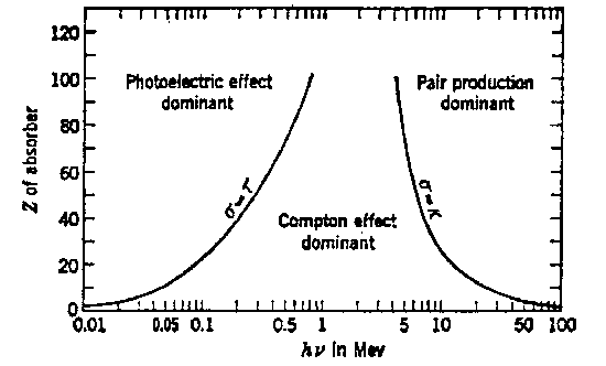
The following figure () (from Evans, p. 712) provides a guide to the relative importance of these three principal interactions over a wide range of energy, hn, of the incident photons and atomic numbers, Z of the attenuating material.
Knowledge of these interactions will also be important in understanding the detection of gammas with a scintillation detector.
Gamma rays may be detected in a number of ways, including gas-filled counters, sold-state detectors, and scintillation detectors. This experiment makes us of a scintillation detector.
In a scintillation detector the gamma ray passes into an organic or inorganic crystal where it interacts with the atoms of the crystal by one of the above interactions. The result of these interactions are the production of charged particles (electrons and gammas) and scattered gammas. The scattered gammas travel a distance through the crystal which depends on their energy and they may or may not interact again before leaving the crystal. The charged particles travel a relatively short distance through the crystal leaving behind a trail of exited atoms. A few materials will emit light in the UV and visible wave lengths as the excited atoms return to the ground state. These ``scintillations'' are detected by a photomultiplier attached to the crystal. Materials to be used as scintillators must of course be transparent to their own light so that it can be seen by the photomultiplier. A discussion of organic and inorganic materials suitable for scintillators is given in an appendix. In this experiment we use an inorganic crystal, NaI, as a scintillator.
If monoenergetic gammas interact in the NaI crystal the pulses produced by the photomultiplier will not all be if the same height but will show a certain distribution in height. A ``pulse height spectrum'' is obtained by plotting a graph of the number of pulses per unit time versus pulse height. The feature of these pulse height spectra can be understood in terms of the interaction the gamma undergoes in the crystal.
In the photoelectric effect and pair production all of the gamma ray energy in converted to electron (or positron) energy. Path lengths of charged particles are short so there is a good chance for all of the energy being absorbed in the crystal. In Compton scattering, however, part of the energy is converted to electron energy and part remains in the scattered photon. The scattered photon may interact again in the crystal by a second Compton scattering or by photoelectric absorption, but in some cases the scattered gamma ray escapes the crystal, carrying its energy with it.
If all the gamma ray energy is captured in the crystal there will still be a pulse height distribution of finite width due to a number of effects which depend on the location of the events in the crystal:
Variations in reflections from the walls.
Variations in photocell sensitivity.
Statistical fluctuation due to the moderate number of photoelectrons in the
photomultiplier.
The pulse height spectrum in this case would look like:
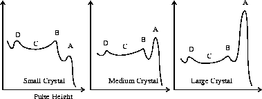
The peak A corresponds to interactions in which no gamma escapes. The peak B and the plateau C correspond to Compton interactions in which the scattered gamma escapes the crystal. B is called the ``Compton Edge'' and corresponds to a Compton scattering in which the gamma is scattered in the backward direction and then escapes the crystal. In this case the photon transfers a maximum amount of energy to the electron so that a minimum of energy escapes. Small peaks and bumps like that at D may be caused in various ways:
All of the above defects (except *) are reduced by using larger (and more expensive) crystals which have a higher probability of both capturing the original gammas and also capturing scattered gammas which might otherwise escape.
The following figure shows a typical spectrum obtained with a
5.1 cm × 5.1 cm
NaI scintillator and a Cs137 source.
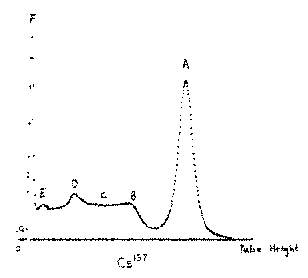
The Cesium-137 nucleus decays by the following scheme:
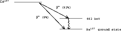
The Cs137 emits an electron (b) plus a neutrino thereby increasing the nuclear charge from 55 to 56 by changing a neutron to a proton.
|
In 92% of the decays only 514 keV is transferred to the electron and neutrino and the Ba137 is left in a metastable, excited state. This decays (T1/2 = 2.6 min.) by either emitting a 662 keV gamma (90%) or ``internally converting'' the gamma (10%) before it leaves the Ba atom and ejecting a K shell electron instead. In effect, the gamma interacts with the K shell electron as the gamma is created by the nucleus. In this case a fast electron is thrown out and then followed by a 32 keV x-ray as the K shell of the Ba is refilled.
The spectrum shows the following features:
|
| Energy (keV) | Source | Lifetime | % Giving Gamma | Origin or Comment |
| 2614.4* | Th228 | 1.9 y | Gives a range of 84 through 2614 keV | |
| 2204.2 | Ra226 | 1600 y | (From daughter Bi214) | |
| 1769.7 | Ra226 | 1600 y | (From daughter Bi214) | |
| 1332.5* | Co60 | 5.3 y | 100 | |
| 1274.5 | Na22 | 2.6 y | 100 | |
| 1173.2* | Co60 | 5.3 y | 100 | |
| 661.6* | Cs137 | 30 y | 85 | |
| 511.0* | Na22 | 2.6 y | 180 | From positron-electron annihilation |
| 356* | Ba133 | 10 y | ||
| 302 | Ba133 | 10 y | ||
| 280* | Se75 | 120 d | 25 | |
| 265 | Se75 | 120 d | 60 | Replace each 2 years |
| 136 | Se75 | 120 d | 57 | |
| 122* | Co57 | 23 d | 98 | |
| 87.7* | Cd109 | 1.3 y | 1.0 | K-X-ray from Ag109 after K capture |
| 81.0* | Ba133 | 10 y | ||
| 75.0 | K-X-ray sometimes from Pb shielding | |||
| 59.5* | Am241 | 458 y | 36 | |
| 32.1* | Cs137 | 30 y | 6.8 | K-X ray from Ba137 after internal conv. |
| 31 | Ba133 | 10 y | K-X-ray from Cs133 after K capture | |
| 22.1 | Cd109 | 1.3 y | 25 | K-X-ray from Ag109 after K capture |
y = year
d = day
The table of available gammas gives 18 energies. However, some are
closer together and since there is little point in using energies within 5% of
each other, you should choose about 13 energies such as those marked *.
A number of radioactive sources (Th228, Ra226, Cs137, Ba133, Cd109, Se75, Co60, Co57 and Na22) are available mounted in small plastic rods and are kept in a lead container in Room 2507. Most of them are supplied by Isotope Products Labs. The sources are not strong (1-100 mCi but they could interfere with the counting in this experiment and for this reason they should be kept inside the shielding when not in use.
The gamma ray detector is a scintillation detector consisting of a scintillation crystal, a photomultiplier tube, and a preamplifier assembled in a single unit supplied by a Mech-Tronics Nuclear. The crystal is a 2 inch diameter, 2 inch thick NaI crystal doped with thallium. The photomultiplier is an RCA 8053 with 10 dynodes and a maximum voltage rating of 1500 volts. The photomultiplier anode output is connected to an amplifier by a short length of coaxial cable.
During operation, the crystal absorbs energy from a gamma event and produces a proportional flash of light. The light flash causes the photomultiplier tube cathode to emit a proportional quantity of electrons. These are attracted from dynode to dynode through the tube with a multiplication effect at each successive dynode due to secondary emission. The highly intensified burst of electrons which arrives at the anode of the tube, still proportional to the energy of origin, is transferred to form a charge at the input capacitor in the preamplifier. The amplifier responds by creating a positive output pulse which retains the basic proportional significance.
There are two connectors on the photomultiplier tube base:
HV: Connects the high voltage power supply to the photomultiplier tube, using HV BNC for the white HV cable.
The amplifier is an ORTEC unit. Usually you will find the model 575A. The amplifier accepts both positive or negative input pulses and provides both pulse shaping and amplitude expansion of these pulses. The amplifier gain is set via a control knob setting and there are both unipolar positive and bipolar positive going outputs on BNC connectors. The function of the amplifier is to produce positive output pulses of suitable amplitude and shape so that they can be fed into the pulse height analyzer.
The multichannel analyzer is a PC containing an ORTEC model 916A pulse height analyzer card (MCA) run by the Maestro II software package. The MCA card contains an Analog to Digital Converter (ADC), single channel analyzer (SCA), multichannel scaler (MCS), and a dual-ported memory. The card, along with the standard software, transforms the personal computer into a multichannel analyzer. An input of 0 to 10 volt positive pulses from a shaping amplifier is the only external signal necessary for pulse height analysis operation. For further details consult the manual for the 916A and the manual for the Maestro II software which runs the MCA board. The software is located in the MCA directory and is started by typing MCA on the command line. The MCA has been set up for the input voltage range of 0 to 10 Volts to correspond to channel 0 (0 V) and channel 512 (10 V).
The high voltage supply for the gamma ray detector is usually a Hewlett Packard Model 6522A using the positive HV output connector. The supply can furnish an output of ± 0 - 2000 V DC. We normallly run the photomultiplier somewhere between 800-1000 Volts.
Any mobile scope may be used to display the pulses being input to the multi-channel pulse height analyzer.
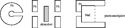
The source is placed in the center of a lead cylinder so that gamma rays may pass freely:
through a second collimating hole in a circular lead plate.
through any absorber.
through a third collimating hole in a second circular lead plate.
through a fourth hole in the lead shielding the NaI detector.
The purpose of the 4 collimating holes is to insure that only gammas which are not scattered may reach the detector. Once a gamma ray has been scattered by the lead or by the absorber, then it is unlikely to reach the detector. The NaI crystal has a diameter of 5.1 cm but the entry hole in the lead has a diameter of only 3.2 cm. This is to insure that the gammas which enter the NaI have a high probability of losing all their energy in the NaI.
Connect the output of the amplifier to the BNC connector labeled ``ADC'' on the MCA card located in the rear of the PC.
Connect the high voltage supply to the ``HV'' connector on the detector using the white HV cable with HV BNC connectors. Turn it on and set to about +800 volts to start.
Place a Cs137 source with a strength near 100 mCi in the cylinder and check the pulses from the preamplifier and amplifier with a scope. Note the amplitude and width of the pulses.
The gamma sources used in this experiment have strengths of up to 100 mCi. Although these are relatively safe sources, you should act as if they are stronger.
Handle the sources by the clear plastic rods.
Report a missing source immediately.
Use the survey meter to check radiation levels around the sources.
Collect and record the spectra from a few other isotopes, such as Na22, Co60, Ba133, and Th228. Always adjust the gain so that the significant peaks are within the range of the analyzer.
Plot the intensity of the 662 keV peak as a function of thickness of A1 on semi-log paper. Draw a straight line through the data points and from the slope of the line determine the linear attenuation coefficient of A1 for 662 keV gammas. Taking the density of Al to be 2.70 g/cm3, calculate the mass attenuation coefficient. Compare your result to that quoted in Melissinos, p. 208.
Using SigmaPlot, fit the above data to a straight line with a least squares fit. Have the computer calculate the slope and the standard deviation of the slope. How does this value of the slope compare with that which you determined graphically?
Calculate the mass attenuation constant, mm, at each energy and plot your results as a function of energy.
Calculate the mass attenuation coefficient for each material and plot your results as a function of the atomic number Z, of the material.
Any indication of pair production.
The Z and E dependence of the photo-electric effect.
Be prepared to answer questions on the following:
The shape of any gamma spectrum.
The principle of operation of a multichannel analyzer.
The principle of operation of the photomultiplier.
The K absorption edge of lead is at 88 keV yet it emits X-rays at only 75 keV.
| Absorber | Z | Density (gm/cm2) | K-shell Edge (KeV) |
| Wax (CH2)n | 2.71 | ||
| Lucite (C5H8O2) | 1.16-1.20 | ||
| Be | 4 | 1.848 | 0.111 |
| C (graphite) | 6 | 1.5 | 0.284 |
| Al | 13 | 2.70 | 1.56 |
| Fe | 26 | 7.86 | 7.114 |
| Ni | 28 | 8.90 | 8.333 |
| Cu | 29 | 8.96 | 8.999 |
| Mo | 42 | 10.2 | 20.00 |
| Sn | 50 | 7.29 | 29.20 |
| W | 74 | 19.1 | 69.52 |
| Au | 79 | 19.3 | 80.72 |
| Hg | 80 | 13.55 | 83.10 |
| Pb | 82 | 11.35 | 88.10 |
| U (depleted 238) | 92 | 18.9 | 115.6 |
Eisberg and Resnick, ``Quantum Physics'' Wiley, (1974).
R.D. Evans ``Gamma Rays'' Chap. 8e in ``American Institute of Physics Handbook'', 3rd Ed., McGraw-Hill, (1972).
Stilbene Crystals.
Polystyrene with traces of Terphenyl and POPOP.
Xylene or Toluene with traces of Terphenyl and POPOP.
Various commercial plastic scintillators such as NE102.
Organic scintillators give fast light pulses - about 5 to 15 nsec long. The basic material usually will absorb the light which it emits and so wavelength shifters are dissolved in the liquid (or plastic) to absorb the short wavelengths and re-emit them with longer wavelengths. The wavelength shifters are usually very complex aromatic substances and are used in very dilute concentrations.
The wavelength shifters also absorb UV (from sunlight or the fluorescent lights) and emit a very pleasant and characteristic blue glow.
Organic scintillators have 3 disadvantages:
Their conversion is not as precise as for NaI. Identical particles giving the same energy loss E in an organic scintillator will result in various light outputs with a ± 10% variation.
When compared with inorganic scintillators such as NaI or CsI, organic scintillators have both lower mass densities and lower average atomic numbers Z. Hence the organic scintillators interact less with gammas (to produce free electrons and ionization) and are usually not used as gamma detectors.
The best are single crystals of ZnS, NaI, CsI or BaF. The light output can be sometimes increased by doping (or ``activating'') the crystal with an element such as thallium. This experiment uses a 5.1cm × 5.1 cm cylinder of NaI with about 0.1% Tl. The Tl increases the light output by about 15%.
Inorganic scintillators give a larger and more precise light pulse than organic scintillators. However, the light pulse is emitted over a longer time, about 1 msec and so NaI is not as useful for fast coincidence work as organic scintillators.
Read Melissinos 194-196.